Diabetic retinopathy is a common complication of diabetes that affects the eyes. It occurs when high blood sugar levels damage the blood vessels in the retina, which is the light-sensitive tissue at the back of the eye that helps us see.
An exam for diabetic retinopathy is important because it can detect changes in the blood vessels of the retina that may indicate the early stages of the condition. Early detection is critical because diabetic retinopathy can lead to vision loss if left untreated.
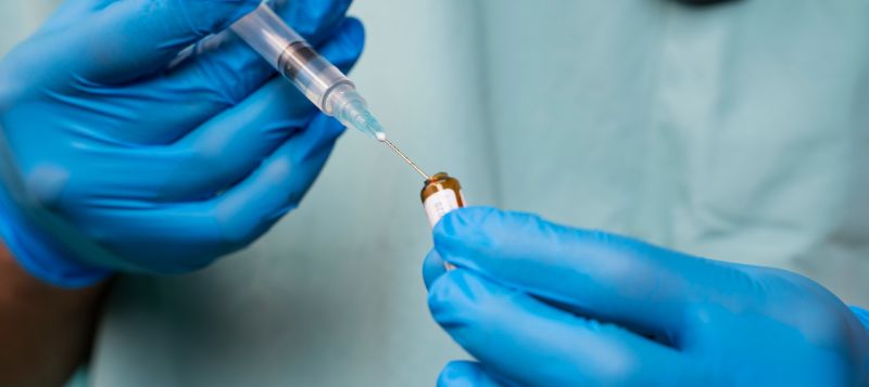
In cases where diabetic retinopathy has progressed to more advanced stages, an intra-vitreal injection of medication may be necessary. This injection delivers medication directly into the vitreous, which is the gel-like substance inside the eye that helps maintain its shape. The medication can help reduce the swelling and inflammation that occur in advanced diabetic retinopathy, which can improve vision and slow the progression of the disease.
Diabetic retinopathy (DR) exams and intra-vitreal injections (IVIs) are both important tests for managing diabetic retinopathy. However, the accuracy of these tests depends on a variety of factors, including the skill and experience of the healthcare professional performing the exam or injection, the quality of the equipment being used, and the patient’s individual factors, such as their age, overall health status, and severity of the disease.
In general, DR exams are a reliable and accurate way to detect and monitor diabetic retinopathy. During a DR exam, an ophthalmologist or optometrist will examine the back of the eye using a special instrument called an ophthalmoscope. This allows them to see the retina and look for signs of damage caused by diabetes, such as leaking blood vessels or swelling. If detected early, diabetic retinopathy can often be managed and treated effectively.

